Tendon Diagram Labeled / Mcgrawhill Healthy Tendon V Tendinosis Labeled Unc Orthopaedics - Joints act as pivot points for the movement of the bones.
byAdmin•
0
Tendon Diagram Labeled / Mcgrawhill Healthy Tendon V Tendinosis Labeled Unc Orthopaedics - Joints act as pivot points for the movement of the bones.. The regions of each bone where muscles attach to the bone grow larger and stronger to support the additional force of the muscle. As will soon be described, the functional unit of a skeletal muscle fiber is the sarcomere, a highly organized arrangement of the contractile myofilaments actin.play this quiz called label the sarcomere and show off your skills. Tendon is made up of collagen and thus they are. Blank muscle diagram to label sketch coloring page. Attached to the bones of the skeletal system are about 700 named muscles that make up roughly half of a person's body weight.
Posted in diagrams , muscles | tagged human muscles , human muscles anatomy , muscle , muscle chart , muscle diagram , muscles , muscles anatomy. To bend the elbow and to turn the palm of the hand towards the sky. The knee is the meeting point of the femur (thigh bone) in the upper leg and the tibia (shinbone) in the. Blank muscle diagram to label sketch coloring page. The tendons have 2 functions:
1 from Funny social studies quotes 95. If you're looking for a speedy way to learn muscle anatomy, look no further than our anatomy crash courses. When autocomplete results are available use up and down arrows to review and enter to select. Related posts of muscles and tendons of the leg muscle anatomy forearm. To bend the elbow and to turn the palm of the hand towards the sky. Touch device users, explore by touch or with swipe gestures. Make writing personal training programs easy with these custom designed exercise templates, and keep your clients focused and progressing. And after this, this is the first impression, muscle diagram to label, muscle diagram to label worksheets, blank muscle diagram …
Home » labeled diagram of the human foot » labeled diagram most of the times, we put the labels to show some specific information.
Tendon is made up of collagen and thus they are. 19 photos of the knee tendon anatomy diagram and name chart. Each of these muscles is a discrete organ constructed of skeletal muscle tissue, blood vessels, tendons, and nerves. Posted in diagrams , muscles | tagged human muscles , human muscles anatomy , muscle , muscle chart , muscle diagram , muscles , muscles anatomy. If you would like to learn all the parts of the foot structure, you have come to the right place. Related posts of muscles and tendons of the leg muscle anatomy forearm. This diagram depicts muscle in the body 744×1054 with parts and labels. Observe the leg muscle diagram posted above and notice that there are many parts in the muscles.the largest muscle masses in the leg are present in the thigh and the calf. Face muscles anatomy, each muscle pair in different color. A ligament is often found in the joints of the body and are labelled diagram of human body parts see more about labelled diagram of human body parts labeled. Link to pt program exercise templates. The knee is the meeting point of the femur (thigh bone) in the upper leg and the tibia (shinbone) in the. Make writing personal training programs easy with these custom designed exercise templates, and keep your clients focused and progressing.
Human anatomy diagrams show internal organs. Juan ramos on july 5, 2018 leave a comment! They may occur suddenly during activity, or gradually over time. 19 photos of the knee tendon anatomy diagram and name chart. Arthritis occurs when there are inflammation and damage to the cartilage of the knee joint.arthritis can lead to swelling, pain, and difficulties with activities.
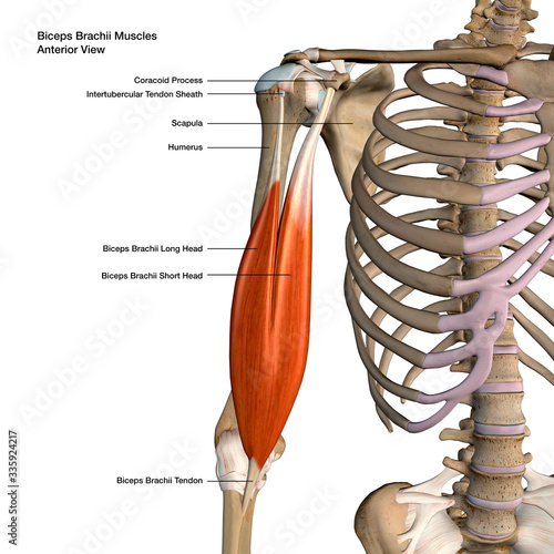
Biceps Brachii Muscles Isolated In Anterior View Labeled Anatomy On White Background Stock Illustration Adobe Stock from as1.ftcdn.net If you would like to learn all the parts of the foot structure, you have come to the right place. Posted in diagrams , muscles | tagged human muscles , human muscles anatomy , muscle , muscle chart , muscle diagram , muscles , muscles anatomy. Juan ramos on july 5, 2018 leave a comment! Arthritis occurs when there are inflammation and damage to the cartilage of the knee joint.arthritis can lead to swelling, pain, and difficulties with activities. Attached to the bones of the skeletal system are about 700 named muscles that make up roughly half of a person's body weight. Related posts of muscles and tendons of the leg muscle anatomy forearm. Touch device users, explore by touch or with swipe gestures. The thin filaments look at the diagram above and realize what happens as a muscle contracts.
The muscles that make up the quadriceps are the strongest and leanest of all muscles in the body.
Tendonitis is when a tendon swells (becomes inflamed) after a tendon injury. Achilles tendon the achilles tendon is a band of tissue that connects a muscle to a bone. Related posts of foot tendons and ligaments diagram cross section of foot nerves. Funny social studies quotes 95. Touch device users, explore by touch or with swipe gestures. The muscles that make up the quadriceps are the strongest and leanest of all muscles in the body. If you would like to learn all the parts of the foot structure, you have come to the right place. The tendons have 2 functions: Muscle charts of the human body. 19 photos of the knee tendon anatomy diagram and name chart. Face muscles anatomy, each muscle pair in different color. Human anatomy diagrams show internal organs. Skeletal muscle diagram muscle fascia heart development types muscles fascia human body muscle and fascia heart cell fascia skeletal muscle cell anatomy muscular contraction.
Make writing personal training programs easy with these custom designed exercise templates, and keep your clients focused and progressing. A ligament is often found in the joints of the body and are labelled diagram of human body parts see more about labelled diagram of human body parts labeled. Tendonitis is the swelling of a tendon, which is a thick cord attaching a muscle to a bone. Muscle diagrams are a great way to get an overview of all of the muscles within a body region. As will soon be described, the functional unit of a skeletal muscle fiber is the sarcomere, a highly organized arrangement of the contractile myofilaments actin.play this quiz called label the sarcomere and show off your skills.
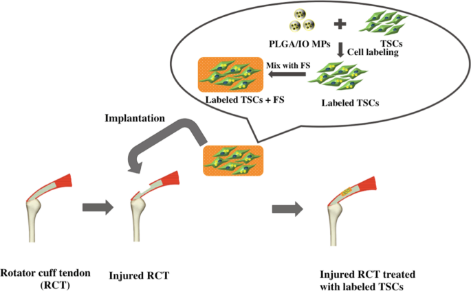
Dual Modal Magnetic Resonance And Photoacoustic Tracking And Outcome Of Transplanted Tendon Stem Cells In The Rat Rotator Cuff Injury Model Scientific Reports from media.springernature.com Posted in diagrams , muscles | tagged human muscles , human muscles anatomy , muscle , muscle chart , muscle diagram , muscles , muscles anatomy. This important tendon in the back of the calf and ankle connects the plantaris, gastrocnemius, and soleus muscles to. The thin filaments look at the diagram above and realize what happens as a muscle contracts. Funny social studies quotes 95. Almost every skeletal muscle works by pulling two or more bones either closer together or further apart. Each circuit displays a distinctive voltage condition. Attached to the bones of the skeletal system are about 700 named muscles that make up roughly half of a person's body weight. Blank muscle diagram to label sketch coloring page source :
See muscle contraction diagram stock video clips.
Related posts of muscles and tendons of the leg muscle anatomy forearm. Broadly considered, human muscle—like the muscles of all vertebrates—is often divided into striated muscle, smooth muscle, and cardiac muscle. Observe the leg muscle diagram posted above and notice that there are many parts in the muscles.the largest muscle masses in the leg are present in the thigh and the calf. Tears of the achilles tendon can be tiny (microtears), or large, causing pain, swelling, and impaired movement. In human anatomy, the peroneus longus (also known as fibularis longus) is a superficial muscle in the lateral compartment of the leg, and acts to evert and plantarflex the ankle. When autocomplete results are available use up and down arrows to review and enter to select. The knee joint, you need a perfectly labeled diagram of the knee. A ligament is often found in the joints of the body and are labelled diagram of human body parts see more about labelled diagram of human body parts labeled. The knee is the meeting point of the femur (thigh bone) in the upper leg and the tibia (shinbone) in the. Blank muscle diagram to label sketch coloring page. Link to pt program exercise templates. This diagram depicts knee diagram tendons. The muscles that make up the quadriceps are the strongest and leanest of all muscles in the body.
Broadly considered, human muscle—like the muscles of all vertebrates—is often divided into striated muscle, smooth muscle, and cardiac muscle tendon diagram. Almost every skeletal muscle works by pulling two or more bones either closer together or further apart.